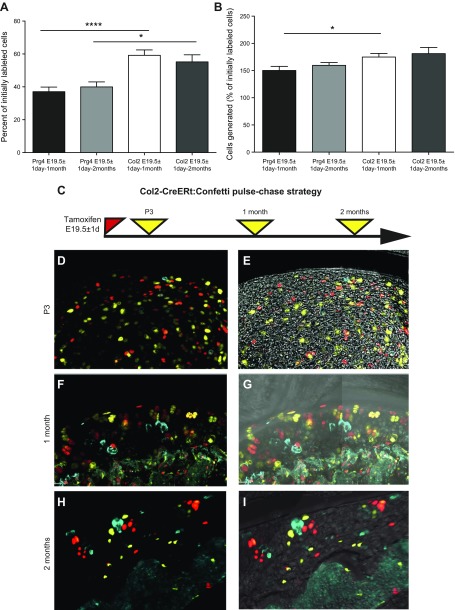
Figure 5.
Assessment of proliferative activity of chondrocytes and superficial cells by clonal tracing. Every single-labeled cell after tracing period indicated was considered as quiescent, and every spatially separated clone that contained cells of the same color counted as initially labeled single progenitor. Thus, the sum of these two gives the number of cells labeled initially. A) The proportion of cells that had proliferated in Prg4-CreER(T2):Confetti mice and Col2-CreERt:Confetti animals traced for 1 or 2 mo was calculated by dividing the number of clones by the number of cells labeled initially. B) Overall increase in cell number was calculated as the total number of labeled cells divided by the number of cells labeled initially. A low degree of recombination (to avoid clonal overlap) was achieved by injecting a smaller amount of tamoxifen. 3D confocal scans of 150-μm–thick sections were analyzed. C) Strategy for labeling Col2-CreER(T):Confetti mice. D–I) Col2-CreER(T)–labeled chondrocytes throughout the articular cartilage, with occasional labeling of superficial cells (D, E). Representative images showing chondrocytes labeled around birth and analyzed 3 d (D, E), 1 mo (F, G), or 2 mo later (H, I; maximum intensity projection shown). On P3, single-labeled cells and clonal doublets were only observed, whereas at 1- and 2-mo timepoints, triplets and quadruplets could be observed. Values are presented as means ± sem; n = 9 mice for every point (A, B). *P < 0.05; ***P < 0.001.

