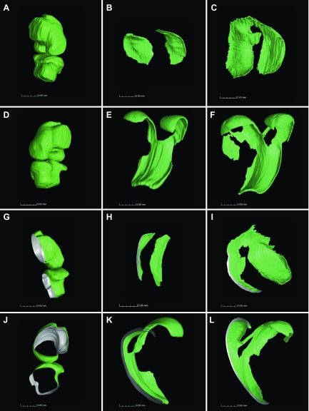
Figure 7.
Visualization of cartilage shape revealed substantial flattening and reshaping in ontogenesis. Cartilage was reconstructed employing VG Studio Max 2.2 software for 3-d-old mouse knee cartilage (A, D, G, J), 1-mo-old tibia (B, H), and 1-mo-old femur (E, K), as well as 2-mo-old tibia (C, I) and femur (F, L). Panels A and D represent different projections of the same joint, G–L cross-sections of CT scans (gray color, section plan), J shows middle sagittal slice of 3-d-old knee, and A–F are the same magnitudes, as well as G–L.

