Figure 1.
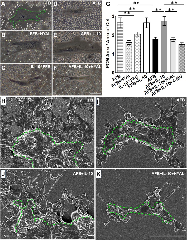
IL-10 induces a fetal-type HA-enriched PCM in AFBs. A–F) Living fibroblasts were imaged with phase contrast to visualize cell bodies, and the area occupied by the pericellular HA coat after 24 h of treatment was assessed by the exclusion of red blood cells from the cell perimeter. White and green dotted lines were artificially drawn in A to delineate the margin of the HA coat and cell body, respectively. Representative cells are shown for FFBs and AFBs under different treatment conditions: FFBs (A), FFBs treated with HYAL (B), IL-10−/− FFBs (C), AFBs (D), AFBs treated with IL-10 (E), and AFBs treated with IL-10 and HYAL (F). G) We quantified the ratio of the means ± sd of 3 experiments (20 cells analyzed per experiment). H–K) Scanning electron microscopy was performed to demonstrate HA cable-like structures on the cell body outlined by green dotted line. Representative cells are shown for different cell types under different treatment conditions: FFBs (H), AFBs (I), AFBs treated with IL-10 (J), and AFBs treated with IL-10 and HYAL (K). See also Supplemental Fig. S1. Scale bar, 50 μm (A–F), 5 μm (H–K). **P < 0.01 by ANOVA and post hoc Bonferroni tests.

