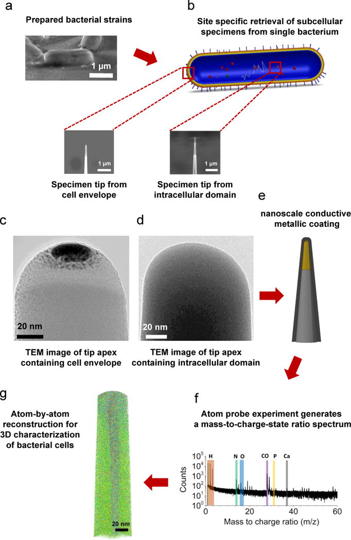
Figure 1.

Schematic of the proposed approach for site-specific 3D atomic-scale analysis of biological cells. (a) Specimen from a single bacterial cells was retrieved using FIB-lift-out technique, and (b) site specific final needle-shaped specimen tip was achieved by precise annular FIB milling and contained a specific region of the original cell (either cell envelope or intracellular domain). (c–d) Selected site specific needle-shaped specimens from the cell envelope and intracellular domains were observed with conventional TEM. (e) Prior to APT, sufficient electrical conductivity is achieved with a nanoscale layer of metallic coating, which allows field evaporation of the specimen tip via pulsed-voltage APT and leads to (f) a mass-to-charge-state ratio spectrum of the ionic species followed by (g) an actual 3D reconstruction of the tomographic map at near-atomic resolution.
