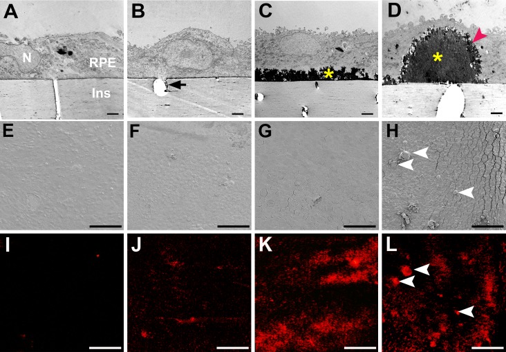
Figure 4.
Evolution of mineralized deposits under porcine RPE in culture. Transmission electron microscopy (A–D), scanning electron microscopy (E–H), and microcomputed tomography for minerals (I–L). (A, E, I) Deposit-incapable, passaged cells at 6 weeks exhibited little or no mineralization. N, nucleus; Ins, culture dish insert. (B, F, J) Deposit-capable primary cells at 6 weeks produced a thin electron-dense sub-RPE deposit that was continuous with material in pores (arrow) and exhibited some mineralization. Due to the sectioning plane, the pore does not cross the Transwell. (C, G, K) At 12 weeks, deposit-capable cells developed a continuous layer of deposit with increased mineralization. (D, H, L) At 26.5 weeks, focal dome-shaped deposits were also present, with diffuse deposit, exhibited intense mineralization (H, L; arrowheads). Diffuse and focal deposits had solid cores (yellow asterisks) with feathery surfaces (red arrowhead) (C, D). Cracks between cells (G, H) are due to brittleness conferred by mineralization. Scale bars denote the following: A–D, 2 μm; E–L, 100 μm.

