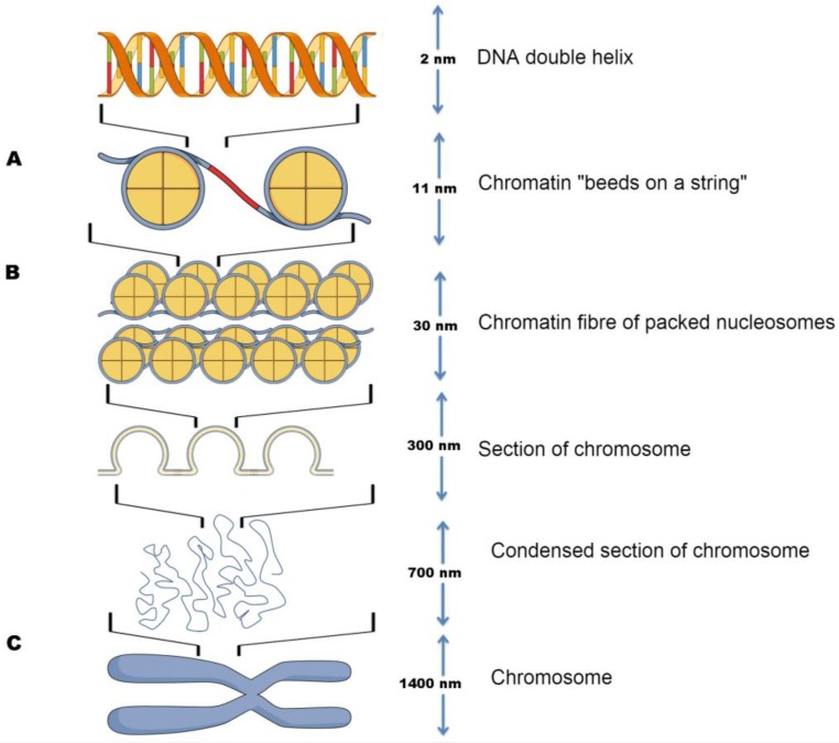
Figure 1.
Schematic of DNA compaction levels facilitated by nucleosomes. Left: (A) DNA wound around the histone octamer, forming a nucleosome; (B) Nucleosomes aggregated into chromatin fibres, which compile into higher order three-dimensional loops and domains; (C) Chromatin fibres assembling into chromosomes. Right: indication of the scale of each successive structures compaction.

