
Extended Data Figure 6.
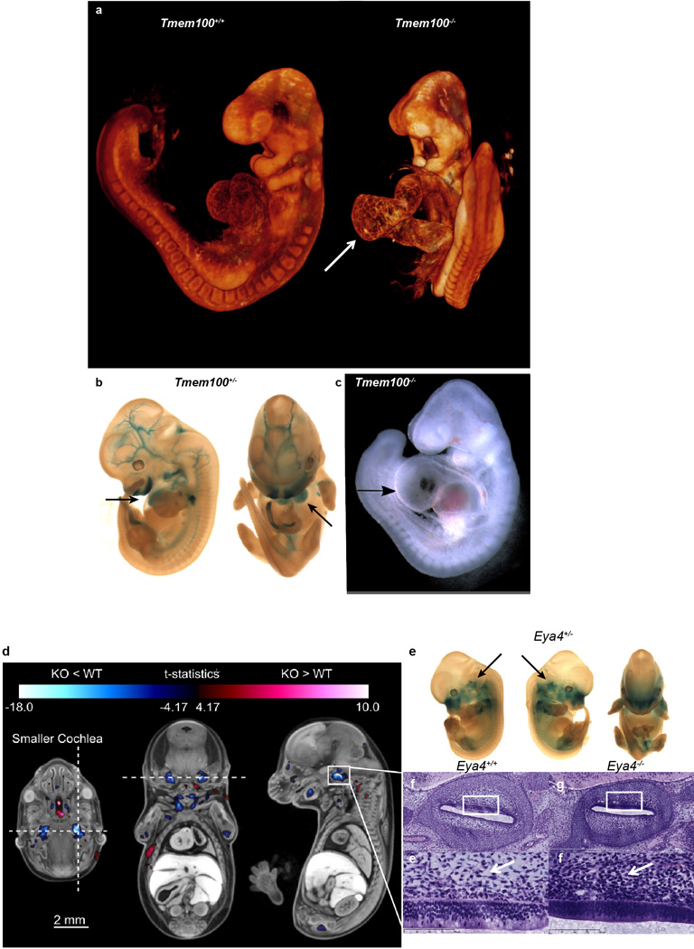
High-resolution 3D imaging reveals phenotypes in Tmem100 and Eya4 mutant embryos. Tmem100−/− embryos have abnormal heart development compared to Tmem100+/+ controls. E9.5 Tmem100−/− embryos had large pericardial effusion and cardiac dysmorphology and enlargement (arrow) when compared to E9.5 Tmem100+/+ (WT) embryos as seen by (a) OPT imaging and (c) bright-field microscopy resulting in lethality. (8 Tmem100+/+ vs. 8 Tmem100−/− with all 8 showing the defect). (b) LacZ expression in the E12.5 Tmem100+/− embryo indicated expression in the heart (arrows), blood vessels and craniofacial regions (blue). d–i, MicroCT imaging revealed a small cochlear volume in E15.5 Eya4−/− embryos. E15.5 Eya4−/− embryos were registered to an average control dataset of the same age followed by automated analysis to show that mutant embryos had a statistically smaller cochlear volume compared to Eya4+/+ (WT) embryos. (d) Transverse, coronal, and sagittal sections through the right cochlea are marked with a horizontal and vertical dashed line in the transverse section to indicate the location of the coronal and sagittal sections, respectively. The color corresponds to areas of larger (red) and smaller (blue) volumes in the KO embryos. The color bar minimum corresponds to a false discovery rate (FDR) threshold of 5%. Hypoplastic bilateral cochlear structures are highlighted in blue. (8 Eya4+/+ (WT) vs. 8 Eya4−/− (KO) with all 8 showing the defect). (e) LacZ imaging in the E12.5 Eya4+/− revealed Eya4 gene expression (blue) in the cochlear region (arrow). (f,g) H&E stained histological sections through the right cochlea of an Eya4+/+ embryo (f) compared to an Eya4−/−embryo (g) confirmed the hypoplastic phenotype. (h,i) Higher magnification of the region (indicated by the white boxes) showed abnormal perilymphatic (periotic) mesenchyme in mutant embryos. In the mutant embryo (i) the perilymphatic mesenchyme did not show rarefaction and had reduced vacuolation versus control (h) (arrows) suggesting the cochlear hypoplasia was due to delayed perilymph development.