
Extended Data Figure 8.
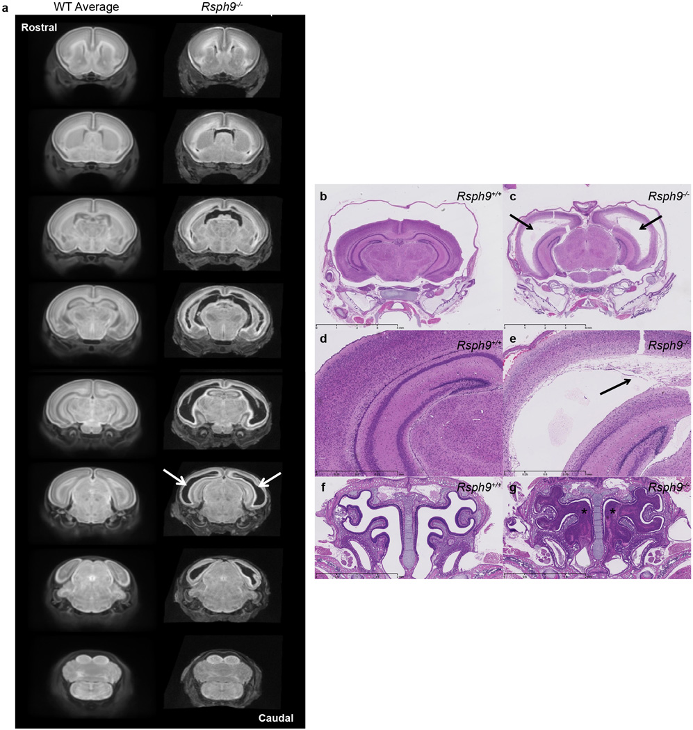
Coronal sections of whole brain MRI revealed enlarged ventricles in the P7 Rsph9−/− (KO) mice. (a) P7 Rsph9−/− mice brains showed enlarged left and right lateral ventricles (arrows) when virtually sectioned from rostral to caudal and compared to a WT average of P7 Rsph9+/+ mice brains. (8 Rsph9+/+ (WT) vs. 10 Rsph9−/− (KO) with all 10 showing the defects). Histological analysis of Rsph9−/− (KO) mice confirmed abnormal brain development. (b,c) Arrows indicate severe hydrocephalus of the left and right lateral ventricles of the Rsph9−/− (KO) P7 mice (c) compared to the Rsph9+/+ (WT) mice (b). The third ventricle was also enlarged but not seen in this section. (d,e) Higher magnification of the cerebrum showed marked rarefaction, cavitation, and loss of periventricular cortical tissue (arrow) in the KO mice (e) compared to WT (d). (f,g) Coronal section through the nasal region revealed that the sinuses of the KO mice were filled with pus (asterisks) (g).