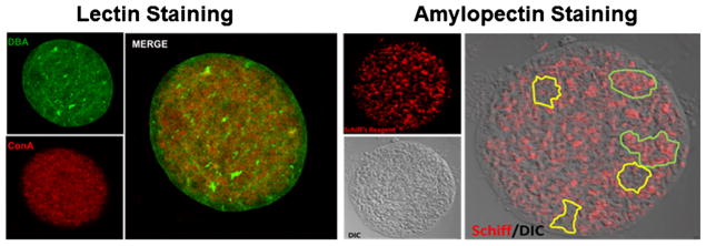
Fig. 1.
Distribution of key glycans in tissue cysts. A distinguishing feature of Toxoplasma gondii tissue cysts is the high level of glycosylation. Left panel lectin staining with FITC-conjugated Dolichos biflorus (DBA-recognizing GalNAc) lectin labels the tissue cyst wall and the intra-cyst matrix (green). In contrast, Concanavalin A (ConA-recognizing mannose and glucose, and an indicator of N-linked glycosylation) selectively stains structures within the bradyzoites and in the matrix (red) but is excluded from the tissue cyst wall (merge). Right panel the distribution of amylopectin granules within bradyzoites detected using Schiff reagent (red) overlaid on a differential interference contrast of a purified tissue cyst reveals an uneven distribution of amylopectin within the tissue cyst with clusters of bradyzoites exhibiting high levels of amylopectin (outlined in green) adjacent to areas with low amylopectin levels (outlined in yellow)

