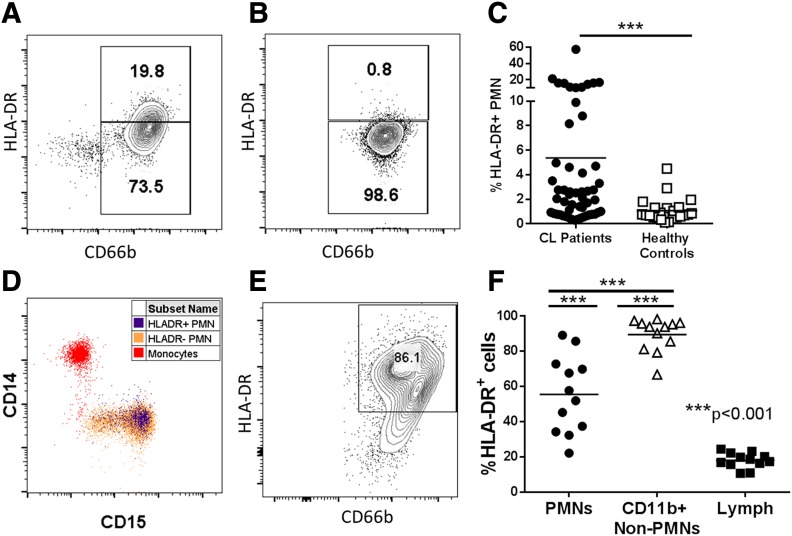
Figure 1. A subset of Brazilian patients with CL has HLA-DR–expressing PMNs in the circulation and cutaneous lesions.
(A and B) Whole blood was isolated from Brazilian patients with CL or endemic healthy controls and stained directly ex vivo for HLA-DR and CD66b. Granulocytes were gated by forward (FSC) and side scatter (SSC) and on single cells resulting in representative flow plots showing HLA-DR expression on CD66b+ neutrophils from a patient with CL and a healthy endemic control, respectively. (C) Quantification of HLA-DR+ expression on total patients vs. endemic controls (unpaired Welch’s t test). (D) Expression of the monocyte marker CD14 and neutrophil marker CD15 is shown on monocytes (red), HLA-DR+ PMNs (blue), or HLA-DR− PMNs (orange). (E) Flow plot showing HLA-DR expression on CD66b+CD15+CD11b+ PMNs from lesions of patients with CL. (F) Quantification of surface HLA-DR on PMNs (CD15+CD66b+CD11b+), non-PMN myeloid cells (CD11b+CD66b−), and lymphocytes (CD11b− FSC, SSC) in lesions from patients with CL.

