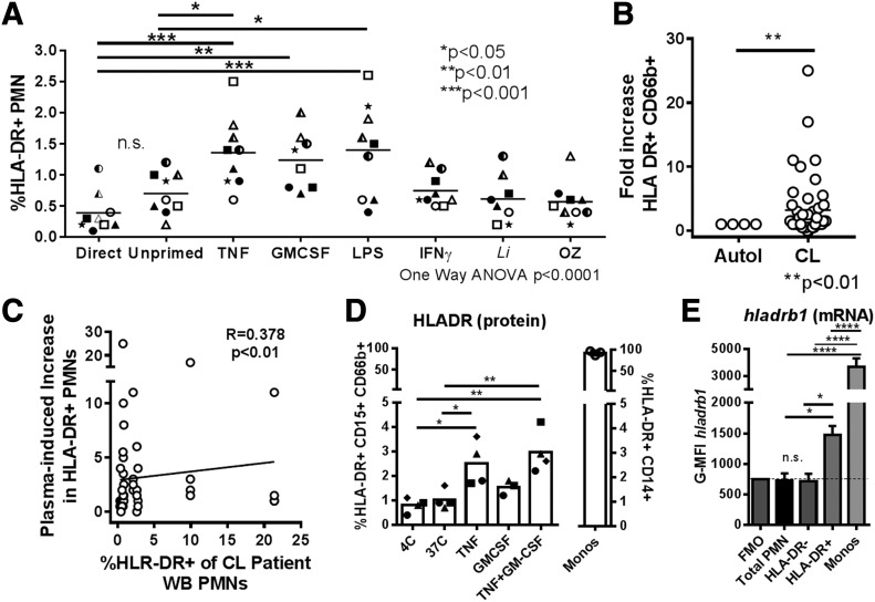
Figure 6. Priming whole blood results in increased surface HLA-DR and hladrb1 mRNA in healthy blood donor PMNs.
(A) Healthy donor whole blood was incubated with priming, inflammatory cytokines, or other stimuli at 37°C. After 2 h, blood was stained for CD66b and HLA-DR. The graph shows the percentage of HLA-DR+ PMNs in plots. Cells from each donor are indicated by unique symbols (statistical analysis: 1ne-way ANOVA with Dunnett’s posttest comparing all conditions to control). (B) Plasma from healthy donor whole blood was removed and replaced with CL patient plasma or autologous plasma. Cells were incubated for 2 h at 37°C, with 5% CO2, followed by staining for surface with HLA-DR on PMNs. (C) Plot showing significant correlation between the expression of HLA-DR on PMNs of patients with CL (x axis) and the increase in HLA-DR seen in healthy donor cells when incubated in that patient’s plasma (y axis). (D and E) After 2 h priming in cytokines, similar to that in panel A, cells were stained for surface HLA-DR (D). The same cells were incubated with a probe hybridizing to intracellular hladrb1 mRNA, which was detected by flow cytometry using PrimeFlow assay (E). The graph shows a plot of the means ± sd MFI corresponding to the hladrb1 mRNA probe in neutrophils staining as HLA-DR− vs. neutrophils staining as HLA-DR+ for the conditions shown in panel D.

