
Figure 1.
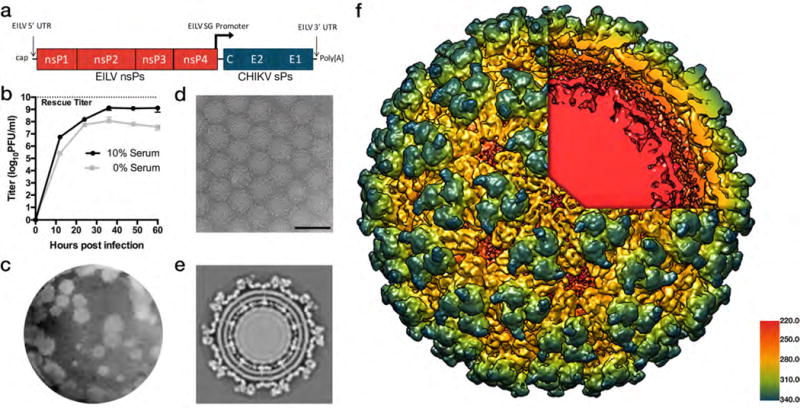
Characterization of EILV/CHIKV. (a) Genome organization of EILV/CHIKV chimeric virus containing the EILV 5′ and 3′ untranslated regions (UTRs) and subgenomic (SG) promoter as well as encoding genes for EILV nonstructural proteins (nsPs) and CHIKV structural proteins (sPs) (b) Replication kinetics of EILV/CHIKV on C7/10 cells in serum-containing or serum-free media. Monolayers were infected at a multiplicity of infection (measured in mosquito cells) of 0.1 PFU/cell and supernatants were collected at the indicated time points and titrated on C7/10 cell monolayers. Each data point represents the mean + s.d. titer of samples from triplicate infections and the experiment was performed twice. (c) EILV/CHIKV plaques 3 days after infection of C7/10 cells. (d) Cryo-electron micrograph (cryoEM) of EILV/CHIKV particles (scale bar = 100 nm). (e) Cross-section of EILV/CHIKV cryo-EM maps revealing transmembrane helices of E2/E1 glycoproteins interacting with capsid proteins. (f) A 9.85Å cryo-EM single-particle reconstruction of EILV/CHIKV, colored radially from E2/E1 heterodimer trimeric spikes in blue, green, and yellow through to the capsid proteins in red underlying the lipid bi-layer revealed through the cutaway. Particle reconstructions were identical to a previously published structure of CHIKV virus-like particles (Supplementary Fig.1).