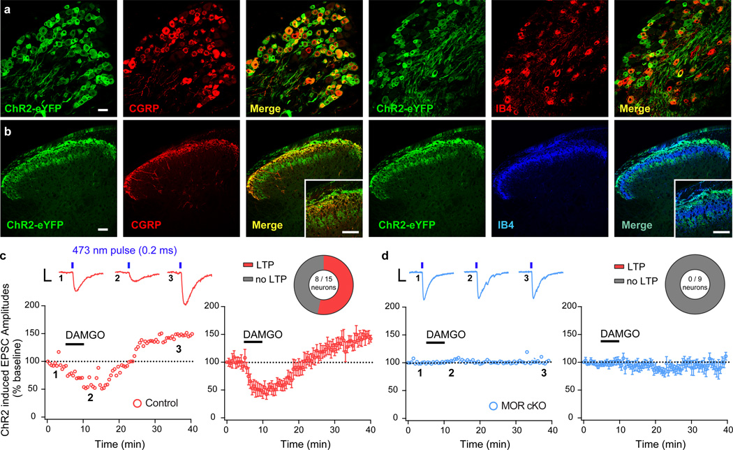
Figure 4. Opioid-induced spinal long-term potentiation (LTP) is initiated by presynaptic MOR in nociceptors.
(a,b) MOR cKO mice crossed with a mouse line expressing Channelrhodopsin2 (ChR2-eYFP) in a Cre-dependent manner produces expression of ChR2-eYFP in Trpv1Cre+ DRG nociceptor cell bodies and central terminals in the spinal cord dorsal horn. Scale bars = 50 µm, throughout. (c,d) Blue light-evoked (473 nm, 1.0 mW/cm2, 0.2 ms, 0.05 Hz) EPSCs recorded in laminae I and II outer spinal neurons in slices from MOR cKO and littermate controls. Numbered inset traces correspond to individual EPSCs before, during, and after wash-out of bath applied DAMGO (500 nM; 5 min duration). Scale bar = 100 pA, 10 ms. (c) DAMGO-induced depression of EPSC amplitude and rebound LTP after washout (Control + LTP; n = 8 / 15 neurons) in control slices. (d) MOR cKO spinal neurons do not show DAMGO-induced depression of light-evoked EPSCs or rebound LTP upon DAMGO washout (n = 9 / 9 neurons). Error bars are mean ± SEM.

