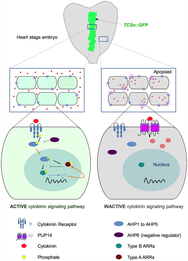
FIGURE 1.
Proposed mechanism of cytokinin-signaling restriction by PUP14 (based on models published by Bürkle et al., 2003; Zürcher et al., 2016). Top: Diagram depicting the region of cytokinin response (indicated in green) revealed by the TCSn:GFP marker in a heart-stage embryo. Zürcher et al. (2016) proposed that PUP14 import of apoplastic cytokinins to the cytosol causes their depletion from the apoplast and makes them unavailable for the membrane-bound AHK receptors that initiate the signaling cascade that activates the TCS response. In the middle and bottom diagrams, cytokinin responsive cells are light green, while non-responsive cells are represented in gray. In the responsive cells (left), cytokinins are perceived by the AHK receptors, which autophosphorylate and subsequently transfer the phosphate (black arrows represent phosphorylation) to Arabidopsis histidine-containing phosphotransfer proteins (AHP1 to AHP5). Then, the AHPs enter the nucleus (orange arrows represent intracellular traffic) and phosphorylate type B Arabidopsis response regulators (ARRs), which activate transcription (blue arrow) of CK response genes, reported by the TCSn:GFP marker line and including type A ARRs (negative regulators of cytokinin signaling, as indicated by blocked lines). In the right part of the diagram, the expression of PUP14 promotes the depletion of cytokinins from the apoplast and the cytokinin response (reported by the TCSn:GFP marker) is not activated, even when exogenous cytokinins are applied. Cytokinins are represented by red circles, and internalized cytokinins are represented by light red circles. For simplicity, only one TCS element or PUP14 transporter per cell is depicted.

