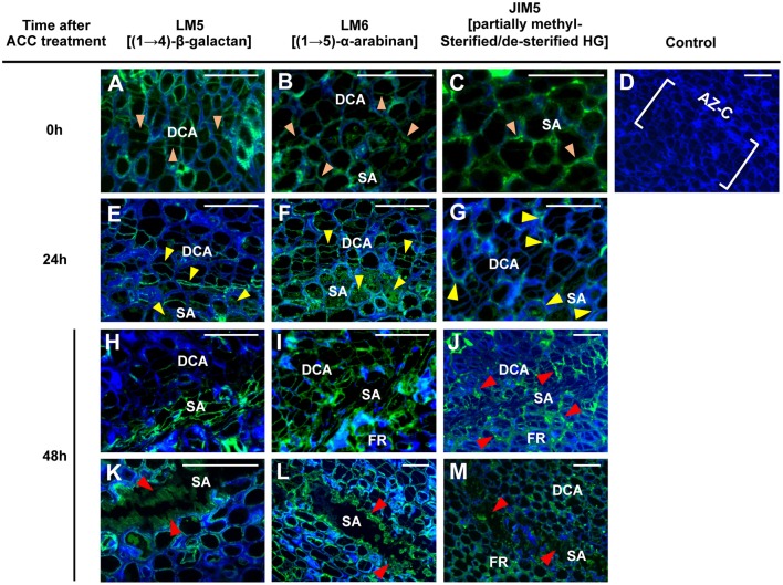
Figure 7.
Immunolocalization of pectic polysaccharides in the AZ-C. Longitudinal sections of tissue containing the AZ-C of Ricalate Navel maturing fruits were incubated with the monoclonal antibodies (mAb) LM5 (A,E,H,K), LM6 (B,F,I,L), and JIM5 (C,G,J,M) to detect (1,4)-β-D-galactans, (1,5)-α-L-arabinans and partially methylesterified/de-esterified HGs, respectively, after 0 (A, B and C), 24 (E,F,G) and 48 h (H–M) of ACC treatment. Control did not show immunofluorescence (D). Scale bars: 5 μm. Key labeling: walls at the two AZ-C cell areas (divided cells area [DCA] and starch-rich area [SA]) and at the fruit rind (FR) cell layers just below the SA showing fluorescence due to each of the mAbs after 0 ( ), 24 (
 ) or 48 (
 ) h of ACC treatment. Micrographs represent the merger of images from pectic epitopes detection by mAbs (green) and from cellulose detection by calcofluor white (blue).

