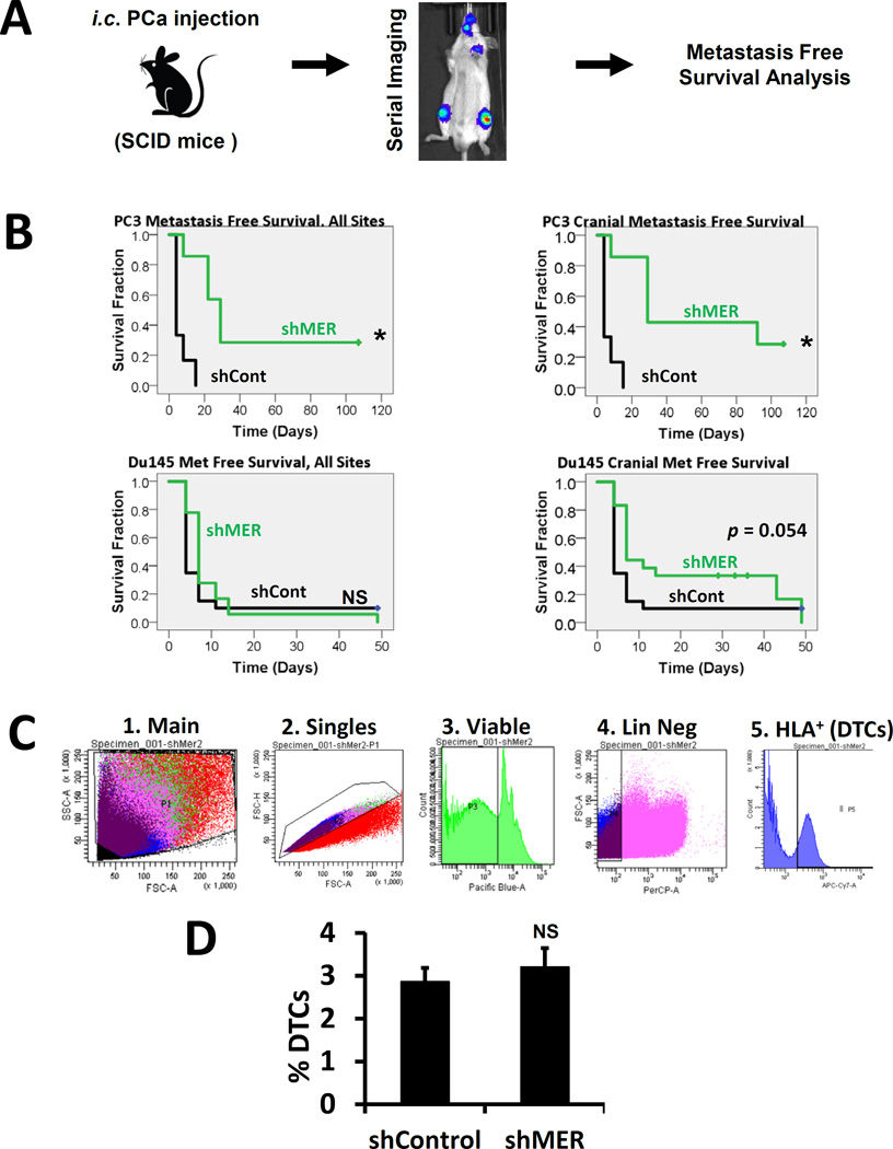
Figure 4.
MERTK knockdown and metastasis free survival in a prostate cancer left ventricle injection xenograft model. A, Experimental design. B, Kaplan-Meier analysis of time to formation of metastases visible by bioluminescence imaging or death in mice injected with luciferase labeled control or shMER PC3 or Du145 cells. Left panels: metastases to any site. Right panels: cranial metastases only.* Indicates p < 0.05 vs control cells. C, Strategy for quantification of the percentage of DTCs in mouse bone marrow by flow cytometry after first depleting the number of mouse cells with immunomagnetic beads. D, Comparison of the percentage of DTCs in mouse bone marrow in control vs. shMER PC3 cells one day after intracardiac injection. Error bars represent mean ± standard error.

