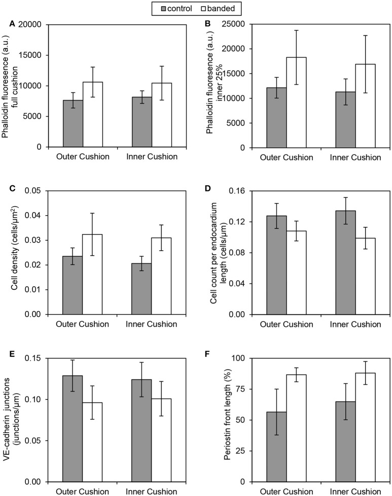
Figure 6.
Quantification comparisons between outer and inner cushions. Phalloidin stain quantifications (A,B), DAPI stain quantifications (C,D), VE-cadherin label quantification (E), and periostin label quantification (F). All control and banded comparisons between outer and inner cushions were not significantly different (p > 0.05).

