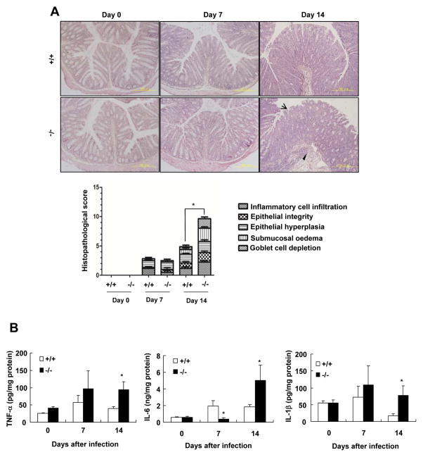
Figure 3. SRC-3−/− mice display more severe colonic pathology compared with wild-type mice after C. rodentium infection.
A, H&E staining of colon slides from uninfected or infected wild-type and SRC-3−/− mice. Arrow heads and arrows denote marked submucosal inflammatory cellular infiltrates/edema and epithelial damage/inflammation in the mucosa, respectively. A 200 × magnification is shown. Histopathalogical scores of wild-type and SRC-3−/− mice at day 0, 7 and 14 after infection. Data represent means + SEM (n=4–10). B, The expression of inflammatory cytokines was higher in the colon of SRC-3−/− mice on day 14 after C. rodentium infection. Data are the means + SEM (n=4–10). *p<0.05. Results are representative of two independent experiments.

