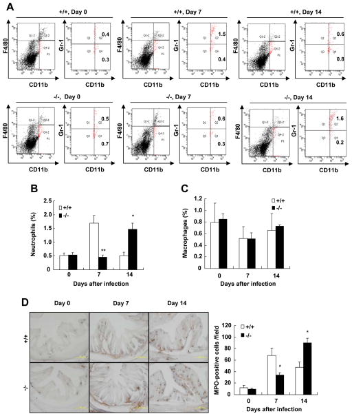
Figure 4. Recruitment of CD11b+F4/80−Gr-1hi neutrophils to the colons is delayed in SRC-3−/− mice after C. rodentium infection.
A, Cells were isolated from the colon of wild-type and SRC-3−/− mice on days 0, 7 and 14 after infection and stained for CD11b, F4/80 and Gr-1 mAb. CD11b+F4/80− cells (total fraction) were gated and percentage of CD11b+F4/80−Gr-1hi cells in the gated population was determined. B, Quantitation of CD11b+F4/80−Gr-1hi neutrophils in the colons of wild-type and SRC-3−/− mice on days 0, 7 and 14 after infection, n=4–5. C, Quantitation of CD11b+F4/80+Gr-1hi inflammatory macrophages in the colons of wild-type and SRC-3−/− mice on days 0, 7 and 14 after infection, n=4–5. D, Examination and quantitation of MPO+ neutrophils. Left panel: Frozen sections of the colon were prepared and stained with Hanker-Yates regent. Brown signal denote MPO+ cells. A 200 × magnification is shown. Right Panel: Quantitation of MPO+ cells. Data are the means + SEM (n=5). *p<0.05; **p<0.01. Results are representative of two independent experiments.

