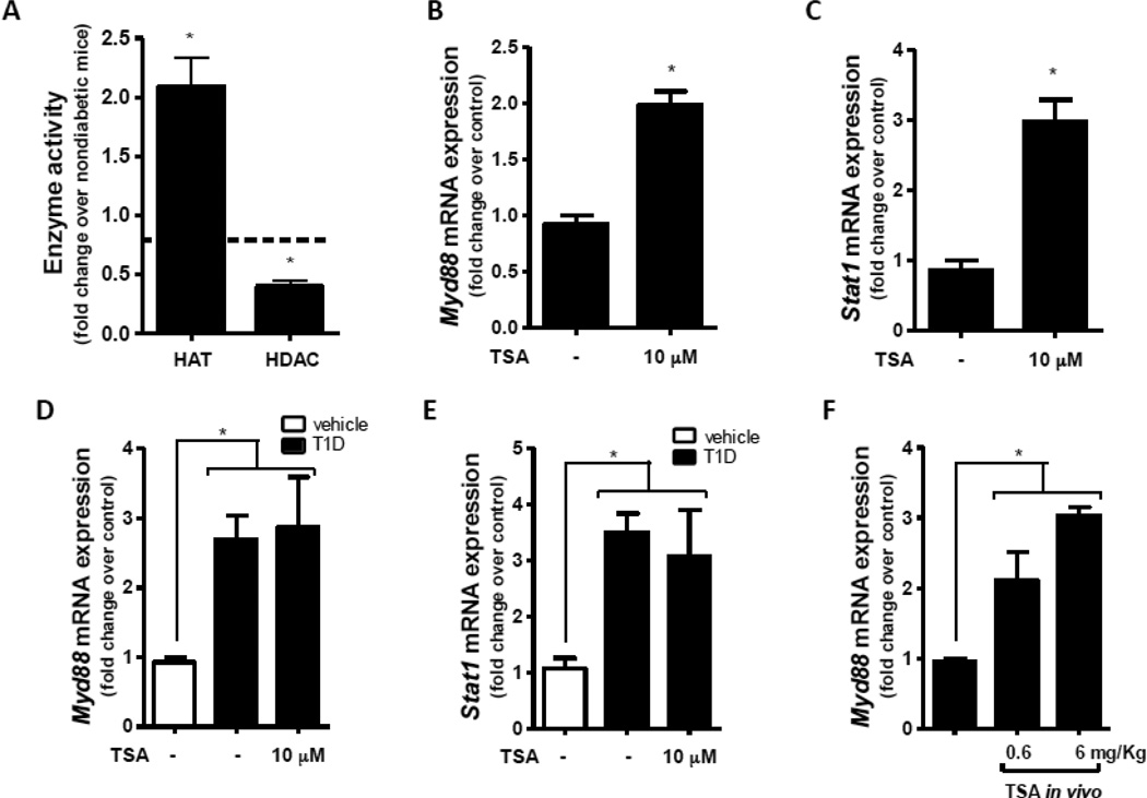
Figure 2.
Activity of HAT and HDAC was analyzed in the nuclear fraction of peritoneal macrophages from T1D and nondiabetic mice as described in Materials and Methods (A). Macrophages from nondiabetic mice were cultured with or without TSA (10 µM) for 24 h, and Myd88 (B) and Stat1 (C) expression were determined by qPCR. Nondiabetic mice (n=4 mice per group) were treated in vivo with TSA (0.6 or 6 mg/Kg) for 24 h; resident peritoneal macrophages were harvested, and Myd88 (D) expression was determined by qPCR. Data are expressed as mean ± SEM from at least 3 independent experiments; *p<0.05 compared to macrophages from nondiabetic mice or compared to peritoneal macrophages isolated from vehicle treated cells.

