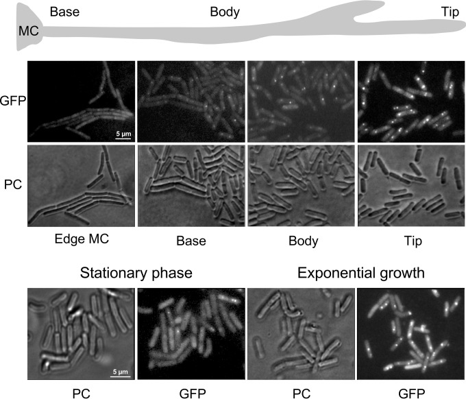
FIG 2 .
Expression and localization of the replication initiator protein DnaA. (Top) Cells from a 1.5-cm swarm producing a fluorescent GFP-DnaA fusion protein (strain SSB2041) were removed from the indicated positions and analyzed by fluorescence microscopy (×100). Panels labeled “PC” show the same cells imaged by phase-contrast microscopy. (Bottom) GFP-DnaA expression in the same strain grown in liquid culture is visualized. Cells were taken either during exponential growth or from the stationary phase and analyzed by fluorescence microscopy under the same conditions as the swarming cells. MC, mother colony.

