
Figure 14.
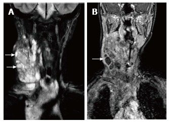
Lymphatic malformation in 13-year-old female patient. Coronal T2WI (A) shows a large, multilocular cystic neck mass with multiple septations. The mass is extending into the superior and anterior mediastinum. Note the variable signal intensity in different locules (arrows). On coronal post contrast T1WI (B), there is enhancement of the wall and internal septations (arrow).