
Figure 17.
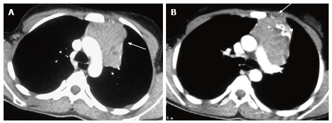
Invasive thymoma in a 13-year-old girl presenting with cough and chest pain. CECT axial images reveal a heterogenously enhancing anterior mediastinal mass with irregular margins (arrow in A) and peripheral calcification (arrowhead in B). There is evidence of focal area of extension to subpleural region anteriorly (arrow in B).