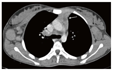
Figure 20.

Tuberculosis in a 12-year-old girl presenting with fever and loss of weight and appetite for 3 mo. CECT axial section reveals heterogenous appearance of the thymus with central low attenuation areas (arrow). There is presence of necrotic right hilar lymph node (arrowhead) and enlarged bilateral axillary lymph nodes (black arrows).
