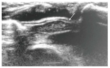
Figure 4.

Normal sonographic (longitudinal view) appearance of the thymus gland in a 2-year-old boy with suspected mediastinal widening on chest radiograph. The thymus is hypoechoic with multiple internal echogenic foci giving the characteristic “starry sky” appearance (arrow).
