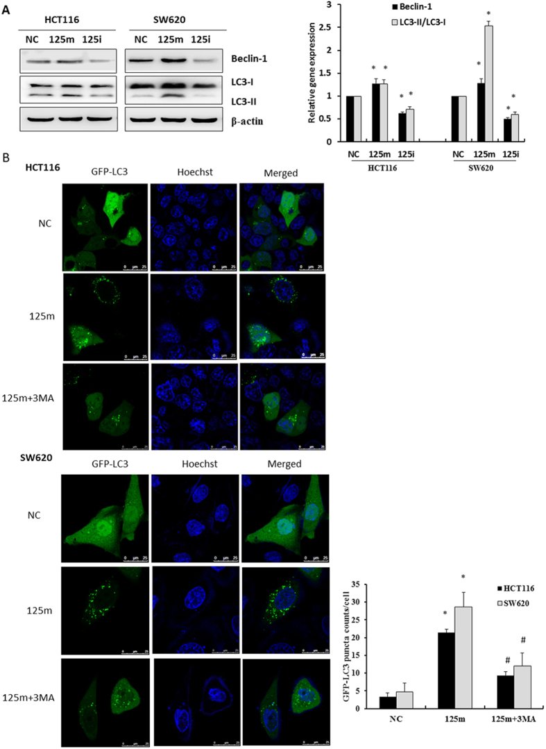
Figure 5. MiR-125b activated autophagic activity in CRC cells.
(A) HCT116 and SW620 cells were transfected with 100 nM miR-125b mimics (125 m) or inhibitors (125i) for 48 h. The expression of beclin-1 and cleaved LC3-II was examined by Western blot assay. Bar graphs indicated relative levels of LC3-II and Beclin-1normalized to β-actin. Data represent mean ± SD of three experiments. *P < 0.05 vs. negative control. (B) HCT116 and SW620 cells were co-transfected with GFP-LC3 plasmid and 100 nM miR-125b mimics for 48 h in the presence or absence of 5 mM 3-MA. Representative photographs were taken using a confocal microscopy (scale bar = 25 μm). Numbers of GFP-LC3 puncta per cell were counted (*P < 0.05 vs. negative control, #P < 0.05 vs. 125 m group, n = 10). Data represent mean ± SD of three experiments.

