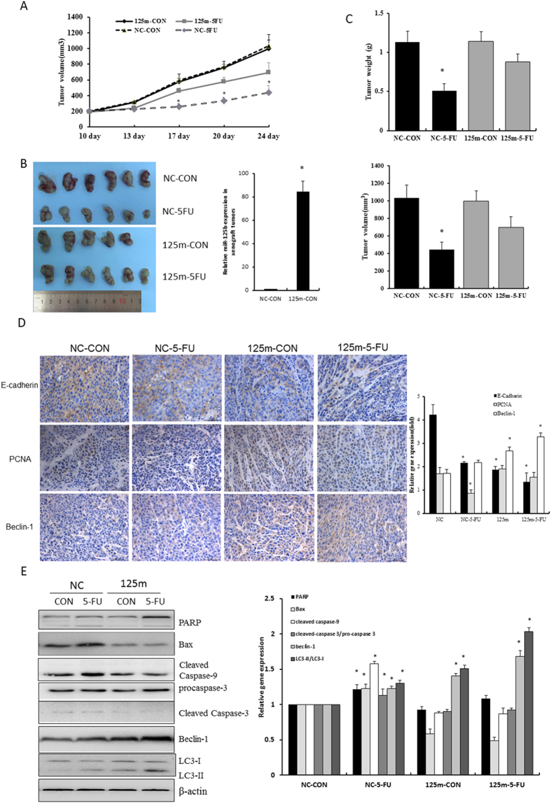
Figure 6. MiR-125b inhibited the 5-FU-induced apoptosis in the xenograft model in vivo.
HCT116 cells infected with lentiviral miR-125b (125 m) and negative control (NC) were injected subcutaneously into nude mice. When tumor volume reached 100 mm3, 5-FU (20 mg/kg) was injected intraperitoneally five times per week for two weeks. (A) Tumor volumes were measured twice per week and the dynamic changes were shown in curves. Data represent mean ± SD. CON represents a short form of vehicle control. (B) Tumor images were taken at the end of the experiments. RNA was extracted from xenograft tumors of NC-CON and 125m-CON groups to determine the expression of miR-125b expression by RT-qPCR assay. (C) Tumor weight and volume were determined after removal. Data represent mean ± SD, *P < 0.05 vs. negative control. (D) Representative images were shown to indicate the expression of E-cadherin, PCNA and Beclin-1 in different groups through immunohistochemistry assay(400 ×). Results were semi-quantified by image-pro plus 6 software and statistical analyses were performed. *P < 0.05 vs. negative control. (E) Protein lysates were extracted from xenograft tumor tissues to determine the expression of Bax, PARP, caspases and autophagic proteins beclin-1 and LC3-II by Western blot assay. Bar graphs indicated the relative levels of cleaved PARP, cleaved caspase-9, caspase-3, LC3-II and Beclin-1 normalized to β-actin. Data represent mean ± SD of three experiments. *P < 0.05 vs. negative control. CON represents a short form of vehicle control, 5-FU is a short form of 5-fluororacil.

