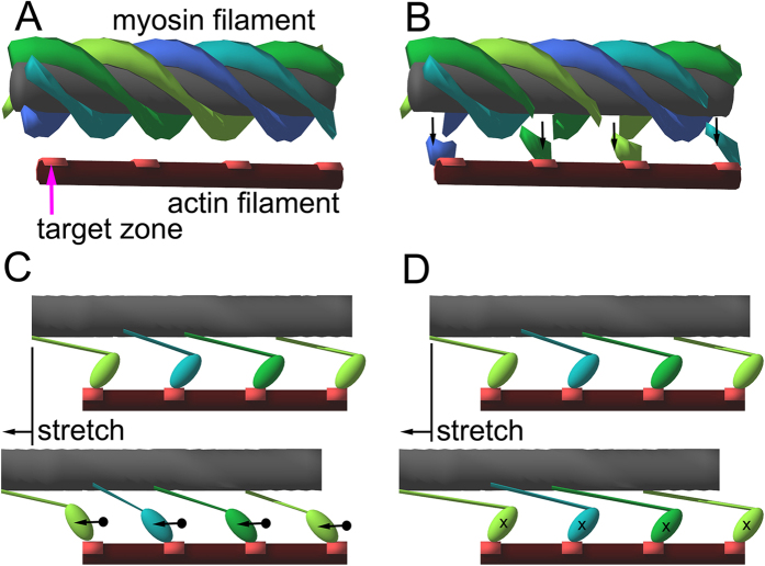
Figure 4. Schematic drawings of the ideas underlying the model calculations.
These drawings summarize structures that can move upon stretch relative to the troponin complexes, causing the change in X-ray interference and resulting in the intensity change of reflections. (A) In the relaxed state. The 4-start helical array of myosin heads (drawn as continuous helical strands) is the only structure that can move with respect to the troponin complexes (located midway between the neighboring actin target zones, not drawn in the figure). (B) Calcium-activated state. The myosin heads close to the target zone leave the helical array (arrows), creating defects in the helical strands. The periodicity of the defects is the same as that of the target zones (or troponin complexes, 38.7 nm) so that these can be sources of interference. (C) Movement of actin-attached myosin heads upon stretch in the weakly binding state. Because of the limited interaction in the actin-myosin interface, a large movement of the center of mass of the myosin head is expected (dots and arrows). (D) Movement of actin-attached myosin heads upon stretch in the strongly binding state. Because of the extensive interaction in the interface, the movement of the center of mass is limited. In (C and D) the myosin heads not bound to actin (helical arrays with defects) are not drawn.

