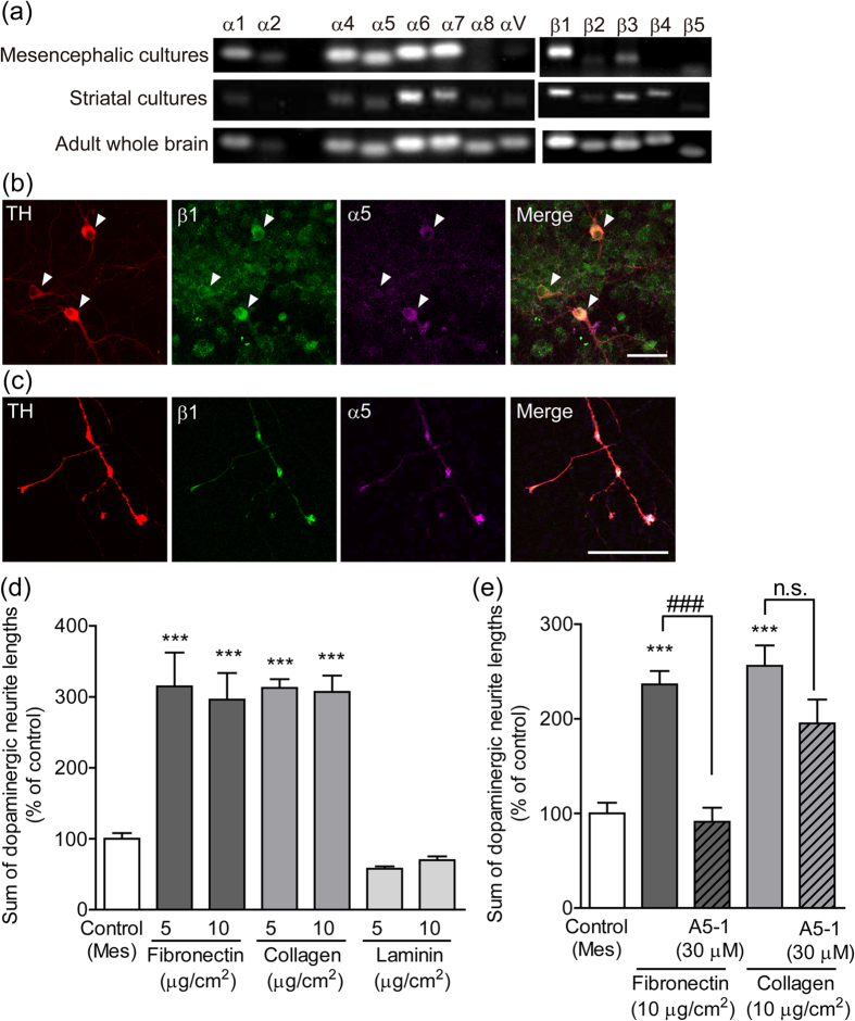
Figure 3. Participation of integrin α5β1 in dopaminergic neurite outgrowth.
(a) Expression of integrin mRNA in mesencephalic and striatal cultures. mRNA levels were analyzed using RT-PCR. (b and c) Expression of integrin α5 and β1 on dopaminergic neurons. Representative photographs of dopaminergic soma (b) and neurites (c). Mesencephalic cells were cultured alone for 10 days and then processed for immunostaining of TH, integrin α5 and integrin β1. Arrowheads indicate dopaminergic soma. Scale bars = 50 μm (b) and 25 μm (c). (d) Influence of the extracellular matrix on dopaminergic neurite outgrowth. The outside of the isolation wall was coated by fibronectin, collagen type I, or laminin (5 and 10 μg/cm2). Mesencephalic cells were cultured alone for 10 days. (e) The effect of integrin α5β1 blocking peptide on dopaminergic neurite outgrowth on the extracellular matrix. The outside of the isolation wall was coated by fibronectin or collagen type I (10 μg/cm2). Mesencephalic cells alone were cultured in the presence or absence of A5-1 (30 μM) for 10 days. ***p < 0.001 vs. control (Mes) group. ###p < 0.001. n.s.: not significant.

