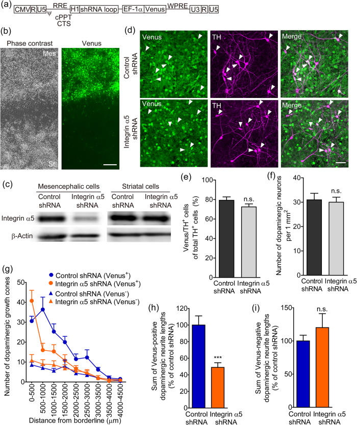
Figure 4. The effect of integrin α5 knockdown in mesencephalic cells on dopaminergic neurite outgrowth to the striatal cell region.
(a) Structure of the lentiviral vector expressing shRNA and Venus under the control of the human H1 and human EF-1α promoters, respectively. (b) Preferential infection of mesencephalic cells with lentiviral vectors. Lentiviral vectors were applied inside the isolation wall during 5 to 24 hours after cell plating. Mesencephalic cells were paired-cultured with striatal cells for 5 days. Representative photographs show the vicinity of the borderline between the cell regions. Left: phase-contrast image. Right: Venus fluorescence. Scale bar = 200 μm. (c) Selective knockdown of integrin α5 in mesencephalic cells. Protein samples were separately collected from the mesencephalic and striatal cells of paired-cultures 6 days after infection with lentiviral vectors. β-Actin was used as a loading control. (d–f) The effects of lentiviral vectors on infection efficiency and cell density in dopaminergic neurons. After infection with lentiviral vectors, mesencephalic cells were paired-cultured with striatal cells for 10 days and then processed for immunostaining of Venus and TH. Arrowheads (d) indicate Venus-positive dopaminergic neurons. Scale bar = 50 μm. n = 20. (g) Distribution of Venus-positive and Venus-negative dopaminergic growth cones from the borderline of the mesencephalic cell region. (h and i) The effect of integrin α5 shRNA infection in mesencephalic cells on dopaminergic neurite outgrowth to the striatal cell region. Venus-positive (h) and Venus-negative (i) dopaminergic neurite lengths were summed. ***p < 0.001 vs. control shRNA. n.s.: not significant.

