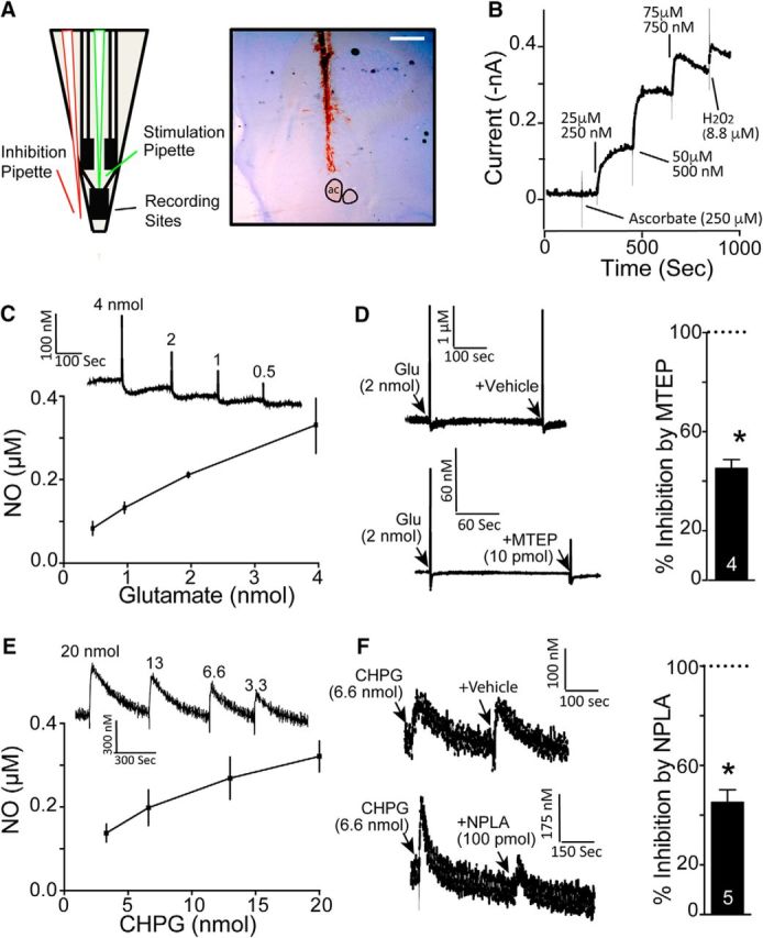
Figure 2.

In vivo electrochemical evidence of glutamate acting via mGluR5 to activate nNOS. A, Schematic of the NO-sensitive electrode, the tip of which was implanted into the NAcore. The glass pipettes were waxed to the electrode assembly such that the tips were ∼100 μm from the surface of the electrode to pressure puff various compounds adjacent to the recording sites. The adjacent cresyl-violet-stained tissue shows an example of electrode placement into NAcore (note blood in electrode track). ac, Anterior commissure. Scale bar, 1 mm. B, In vitro calibration of NO electrodes performed after stabilization of baseline electrodes by measuring the amperometric response at +0.7 V versus Ag/AgCl of a nafion/o-PD microelectrode to AA (interferent), followed by increasing concentrations of DETA/NO (NO donor) and finally to H2O2 (positive control). C, Puffing glutamate adjacent to an NO electrode produced a dose-dependent increase in NO (n = 3). D, The effect of glutamate was antagonized by the mGluR5-negative allosteric modulator MTEP (10 pmol). Left shows example traces and right shows group data normalized to peak amplitude of the glutamate signal in each animal (*p < 0.05). E, Puffing the mGluR5 agonist CHPG produced a dose-dependent increase in NO (n = 3). F, Increase by CHPG (20 nmol) was inhibited by the nNOS antagonist NPLA (100 pmol, *p < 0.05).
