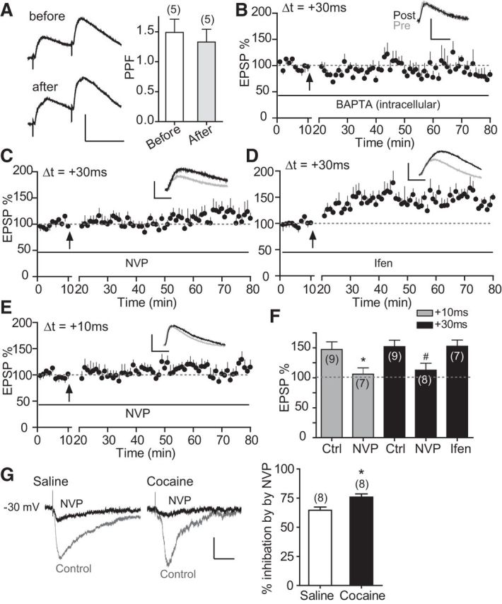
Figure 4.

Cocaine-induced extended-timing t-LTP is expressed postsynaptically and depends on GluN2A-NMDARs. A, PPF before and 30 min after t-LTP induction (60 EPSP–AP pairs, 0.1 Hz, +30 ms). Left, Representative traces with a 50 ms interpulse interval. Scale bars, 2 mV, 50 ms. Right, Summary graph. B, Effect of intracellularly loaded BAPTA (15 mm) on t-LTP induction at +30 ms. C, D, Effects of bath-applied NVP-AAM077 (NVP, 0.4 μm, C) and ifenprodil (Ifen, 3 μm, D) on t-LTP induction at +30 ms. E, Effects of bath-applied NVP-AAM077 (0.4 μm) on t-LTP induction at +10 ms. Scale bars (B–E), 2 mV, 20 ms. Drugs were applied to the extracellular bath during EPSP–AP pairings. F, t-LTP summary. *p < 0.05 versus +10 ms-Ctrl, unpaired two-tailed t test; #p < 0.05 versus +30 ms Ctrl, one-way ANOVA with post hoc Dunnett's test. All experiments in A–F were conducted in slices prepared from repeated cocaine-treated mice. Ctrl t-LTP at +10 and +30 ms in F are replotted from Figure 1 for direct comparisons. G, Repeated cocaine exposure enhances NVP-AAM077-sensitive, GluN2A-mediated NMDAR current. Left, Representative EPSCNMDA recorded at −30 mV in the absence followed by the presence of NVP-AAM077 (0.4 μm) from mice exposed to either saline or cocaine. Right, Summary of NVP-AAM077 inhibition of EPSCNMDA. NVP-AAM077 was applied to the bath to block GluN2A-mediated component after 10–15 min recording of baseline total EPSCNMDA. *p < 0.05, Student's t test. Scale bars, 50 pA, 50 ms.
