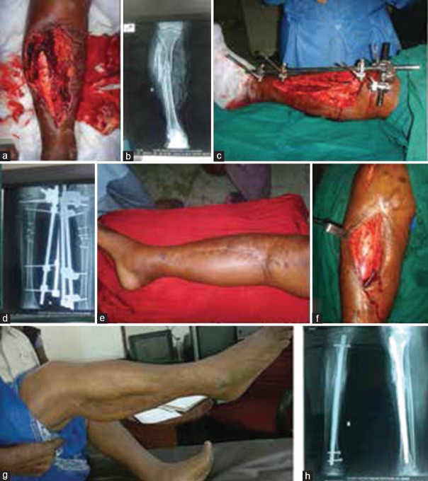
Figure 3.
(a) Clinical photograph (preoperative) of 70 years old lady showing Gustilo Anderson III B open proximal third tibial fracture (b) Preoperative x-ray anteroposterior view showing fracture proximal tibia (c) Clinical photograph showing external fixator in situ with fasciocutaneous skin flap (d) Postoperative X-ray showing external fixator (e) Clinical photograph showing wound healing after removal of external fixator (f) Clinical photograph showing exposure of nonunion site for bone grafting, secondary nailing also done (g) Clinical photograph showing range of motion and healed wounds (h) X-ray anteroposterior and lateral views of leg bones showing fracture union

