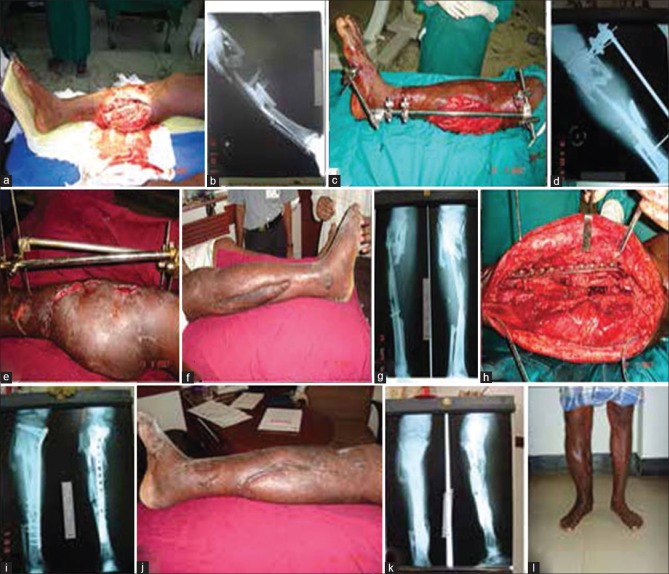
Figure 4.
(a) Clinical photograph (preoperative) of a 45 years old male showing Gustilo Anderson III B middle third tibial fracture (b) X-ray of leg bones anteroposterior view showing fracture middle 1/3rd tibia (c) Clinical photograph showing external fixator in situ with fasciocutaneous skin flap (d) X-ray of leg bones anteroposterior view showing external fixator in situ (e) Clinical photograph showing healing by fascio-septo-cutaneous shift while in external fixator (f) Clinical photograph showing healed wound after removal of external fixator (g) X-ray of leg bones anteroposterior and lateral views after removal of external fixator showing gap nonunion (h) Peroperative photograph showing plating and bone grafting (i) X-ray of leg bones anteroposterior and lateral views showing bone grafting and plating (j) Clinical photograph after plating showing well healed wounds (k) X-ray of leg bones anteroposterior and lateral views showing fracture union after plate removal (l) Clinical photograph in standing position showing healed wounds

