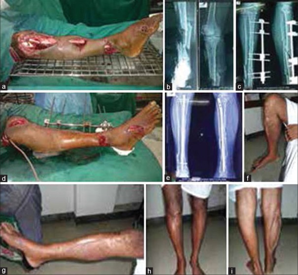
Figure 6.
(a) Clinical photograph (preoperative) of a 20 years old male showing Gustilo Anderson III B segmental tibial fracture (b) Preoperative x-ray of leg bones anteroposterior view showing segmental fracture of tibia (c) X-ray of leg bones anteroposterior and lateral views showing external fixator in situ (d)Clinical photograph showing external fixator in situ (e) X-ray of leg bones anteroposterior and lateral views showing fracture union following secondary nailing (f) Clinical photograph showing flexion of knee (g) Clinical photograph showing extension of knee (h) Clinical photograph showing standing position (anterior) (i) Clinical photograph showing standing position (posterior)

