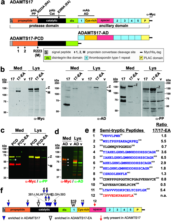
Figure 1. ADAMTS17 undergoes autocatalytic processing.
(a) Domain organization of ADAMTS17 and the constructs ADAMTS17-PCD and ADAMTS17-AD. The location of the ADAMTS17 antibody epitopes (black line) and the sites of site-directed mutagenesis (furin-site: R223A, active site: E390A) are indicated above the ADAMTS17 domains. The predicted furin/PACE cleavage sites are shown below the ADAMTS17-PCD construct. Cleavage at site R223 results in mature (“M”) ADAMTS17. (b) Western blot analysis of conditioned medium (Med) and cell lysate (Lys) from full-length ADAMTS17 (17), the active site mutant ADAMTS17EA (17-EA), and empty vector (v) expressing HEK293F cells. Western blots were probed with the indicated antibodies and detected with enhanced chemiluminescence. (c) Western blot analysis of conditioned medium (Med) and cell lysate (Lys) from ADAMTS17-PCD (PCD), ADAMTS17-PCDEA (PCD-EA), and empty vector (v) expressing HEK293F cells. The asterisk indicates a band which is reactive with anti-propeptide antibody (anti-PP, green), but not anti-myc (red), and which is absent in ADAMTS17-PCDEA. (d) Western blot analysis of conditioned medium (Med) and cell lysate (Lys) from ADAMTS17-AD (AD) and empty vector (v) expressing HEK293F cells. The asterisk indicates intact ADAMTS17-AD (yellow), reactive with anti-myc (red) and anti-ancillary domain antibody (anti-AD, green). Arrowheads indicate species reactive with only anti-myc, but not anti-AD (red). (e) Semi-tryptic peptides resulting from ADAMTS17 autoproteolysis and identified by LC-MS/MS (blue: enriched in ADAMTS17, black: enriched in ADAMTS17EA, red: present only in wild-type ADAMTS17) (f) Location of semi-tryptic peptides in ADAMTS17. Numbers and color coding correspond to the list in e. mAb, monoclonal antibody; M, mature enzyme; pAb, polyclonal antibody; v, vector; Z, zymogen.

