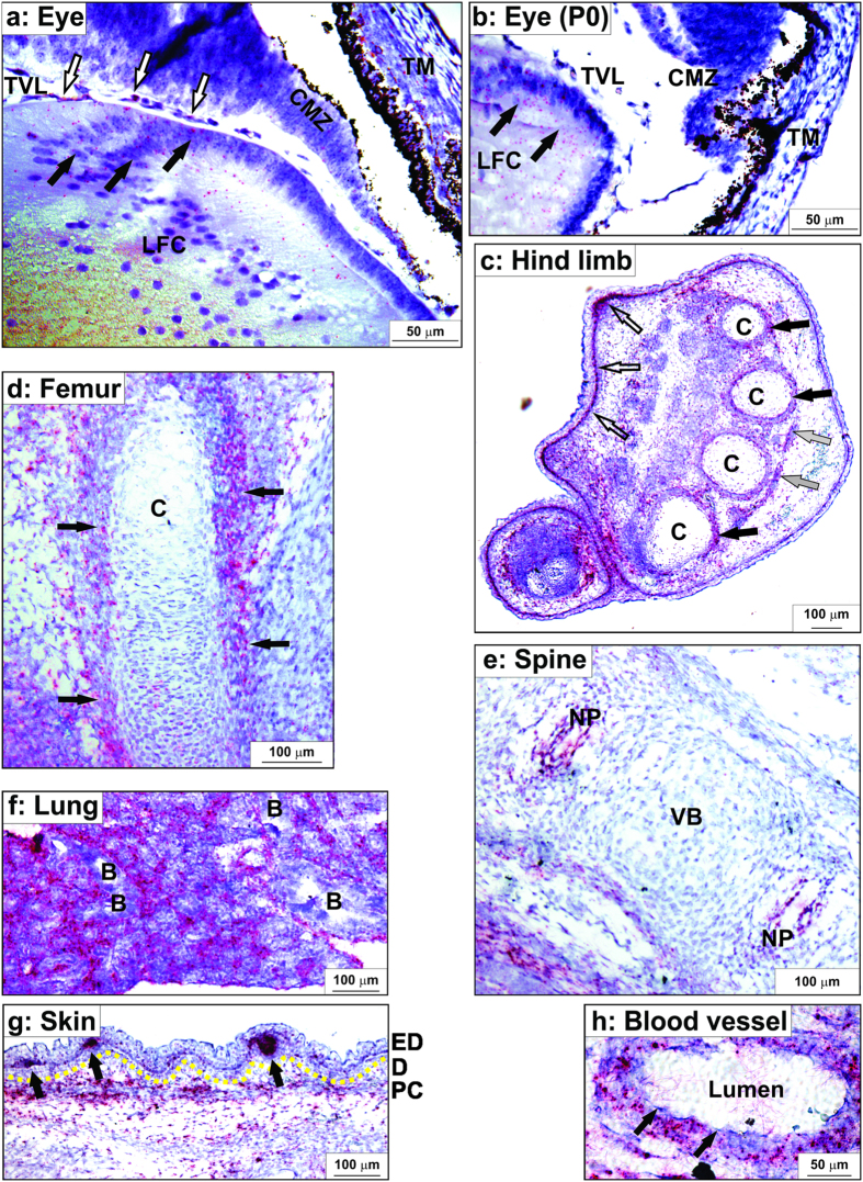
Figure 5. Adamts17 in situ hybridization in 16.5 day-old mouse embryos or neonates showing expression in tissues relevant to Weill-Marchesani syndrome.
The RNAScope method results in red signal over cells containing Adamts17 mRNA. Sections were counterstained with hematoxylin (blue). (a,b) At embryonic day (E) 16.5 (a), Adamts17 was expressed in equatorial lens fiber cells (LFC, black arrows), capillaries of the tunica vasculosa lentis (TVL, white arrows), the non-pigmented epithelium of the ciliary margin zone (CMZ), and the trabecular meshwork cells (TM) of the eye. In neonates (b) Adamts17 continued to be expressed in LFC, but expression was reduced in the other ocular tissues. (c–e) Adamts17 mRNA expression in the skeleton. In the hind limb autopod (c), Adamts17 was detected in the perichondrium around cartilage (C, black arrows), in tendon (grey arrows), and in the dermis and hair follicles (white arrows). Adamts17 was expressed in the perichondrium of the femur (d) (arrows) and in the nucleus pulposus (NP) of the intervertebral disks but not in the vertebral bodies (VB) (e). (f) Strong Adamts17 mRNA expression was found in the lung parenchyma, but not the bronchial epithelium (B). (g) Adamts17 mRNA expression in skin was localized to the hair follicles and the panniculus carnosus (PC). A yellow line marks the dermal (D) – epidermal (ED) junction. Arrows indicate developing hair follicles. (h) In blood vessels, Adamts17 was expressed in the smooth muscle cells of the blood vessel wall, but not endothelial cells (arrows).

