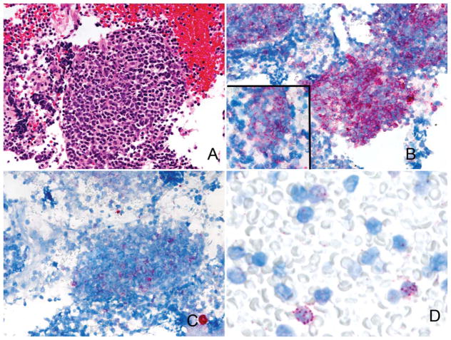
Figure 6.
(A) Peripancreatic lymph node from a patient with a grade 1 follicular lymphoma (H & E). (B) The immunoglobulin κ constant stain is intensely positive, and a follicular pattern is appreciable. The negatively stained cells represent T cells (results not shown). (C) Occasional cells are positive on the immunoglobulin λ constant stain. (D) However, as shown at a high-power magnification, these cells show predominantly nuclear reactivity, a pattern typical of immunoglobulin λ-like polypeptide 5 reactivity (in situ hybridization assay).

