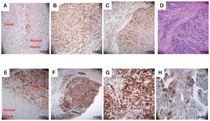
Figure 3.
Representative staining patterns of fibrous septa (FS; A) and tumor lobules (TLs; B and C) of formalin-fixed, paraffin-embedded primary ICC lesions with PD-1–specific mAb. H&E staining of tumor tissue section provides orientation (D). Representative tumor cell staining patterns (E, marginal/interface; F and G, diffuse; H, patchy;) of formalin-fixed, paraffin-embedded primary ICC lesions with PD-L1–specific mAb. Magnification is indicated.

