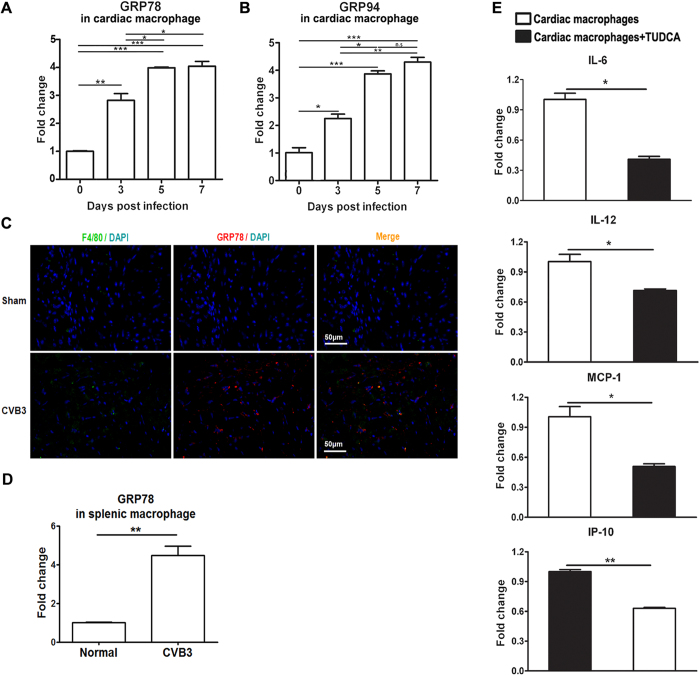
Figure 2. ER stress occurred in the cardiac infiltrating macrophages and facilitated the pro-inflammatory cytokine production.
(A) Dynamic expression of GRP78 in the cardiac macrophages post infection. (B) Dynamic expression of GRP94 in the cardiac macrophages post infection. (C) Immunofluorescence analysis of the induction of ER stress in macrophages derived from CVB3-infected mice. Heart sections from sham-infected mice (upper panels) or CVB3-infected mice (lower panels) were stained with Cy3-labeled rabbit anti-mouse GRP78 (red) or FITC-labeled rat anti-mouse F4/80 (green), and DAPI identifying the nucleus (blue). Images are representative of at least 3 independent determinations. (D) Splenic macrophages were isolated at day 7 post infection and the expression of GRP78 was detected by real-time PCR. (E) Cardiac macrophages were isolated at day 7 post infection and treated with TUDCA, the production of pro-inflammatory cytokines (IL-6, IL-12, MCP-1 and IP-10) was determined by real-time PCR. Each group contained 5–6 mice. Individual experiment was conducted 3 times with similar results. *P < 0.05, **P < 0.01, ***P < 0.001.

