Fig. 7.

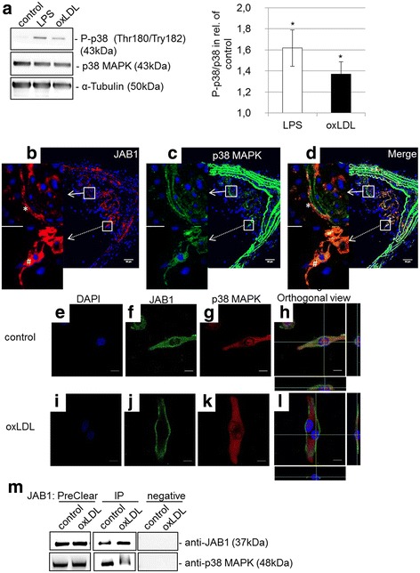
OxLDL-induced phosphorylation of p38 MAPK and interaction with JAB1 in vitro in human MΦ and in arteriosclerotic plaques. a The protein levels of p38 MAPK and P-p38 (Thr180, Try182) were determined by western blot analysis after treatment (4 h) of PMA-differentiated human U937 MΦ with or without 50 μg/ml oxLDL or 0.1 μg/ml LPS. Expression was normalized against α-tubulin. Bars represent mean ± SEM of 4 independent experiments (*p < 0.05). b–d Representative cross-sections of the brachiocephalic trunk of ApoE−/− mice at 30 weeks of age. JAB1 (b) is co-localized (broken arrow) and not co-localized (white arrow) with p38 MAPK (c) in atherosclerotic plaque, as seen in merge (d). JAB1 was detected on the cell surface/cell membrane (*) or in the cytosol (#) in atherosclerotic plaque (b, d). Immunofluorescent staining of JAB1 and p38 MAPK was observed by confocal microscopy (Nikon Eclipse) and analyzed with Fiji ImageJ. The individual channels are depicted in columns: blue, DNA; red, JAB1/COPS5; green, p38 MAPK. Representative photos from 4 independent experiments with similar results are shown. Bars: 50 μm (b, c, d). e–l Immunofluorescence of JAB1 (f, j) and p38 MAPK (g, k) in PMA-differentiated human U937 MΦ was observed by confocal laser scanning microscopy (Nikon Eclipse) and analyzed with Fiji ImageJ. The individual channels are depicted in columns: blue, DNA; green, JAB1/COPS5; red, p38 MAPK. (h, l) The white cross represents the intersection of x-, y- and z-axis in the orthogonal coordinate system. Representative immunofluorescence results from 3 independent experiments are shown. Scale bars: 10 μm. m Immunoprecipitation (IP) of JAB1 was carried out with anti-JAB1 [2A10.8] (GenTex) antibodies followed by immunoblot detection for JAB1 and p38 MAPK in PreClear cell lysates. The eluate from Control Agarose Resin served as negative control. The blot is representative for 4 independent experiments
