Fig. 6.

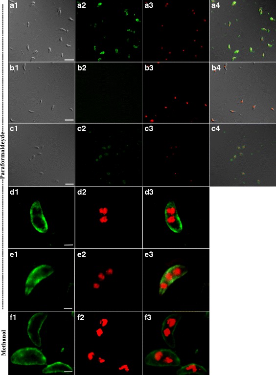
Localization of CSUI_005805 antigens in merozoites and sporozoites. a–c Wide-field epifluorescence microscope (×63 magnification; scale-bar: 10 μm). d–f Meta confocal laser scanning microscope (×63 magnification; scale-bar: 5 μm). a C. suis merozoites: (a1) Differential interference contrast (DIC); (a2) Localization using A488; (a3) Nuclear staining with DAPI (a4) DIC, A488 and DAPI merged. b Negative control, merozoites probed with negative chicken serum: (b1) DIC; (b2) A488; (b3) DAPI; (b4) merged. c C. suis sporozoites: (c1) DIC; (c2) A488; (c3) DAPI; (c4) merged. d and e Paraformaldehyde-fixed C. suis merozoites: (d1, e1) A488; (d2, e2) DAPI; (d3, e3) merged. f Methanol-fixed C. suis merozoites: (f1) A488; (f2) DAPI; (f3) merged
