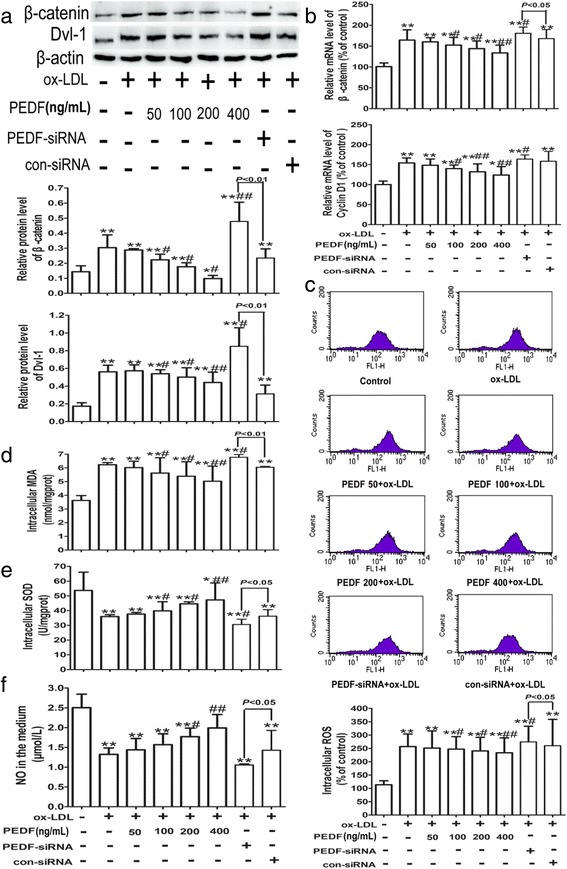
Fig. 6.

PEDF inhibits ox-LDL-induced Wnt/β-catenin pathway activation and oxidative stress in HUVECs. Cells were treated as described in Fig. 5. The protein (a) or mRNA (b) levels of β-catenin, Dvl-1 and Cyclin D1 in HUVECs were analyzed by western blotting and quantitative real-time PCR, respectively. Generation of intracellular ROS (c) was detected by flow cytometry using DCFH-DA as the substrate. Intracellular MDA content (d), SOD activity (e) and NO level (f) in the medium were tested with commercially available kits with a microplate reader according to the manufacturer’s instructions. All data are expressed as the mean ± SD of at least 3 independent experiments. *P < 0.05, **P <0.01 versus the control group; # P < 0.05, ## P < 0.01 versus the 100 mg/L ox-LDL group. β-catenin, non-phosphorylated-β-catenin
