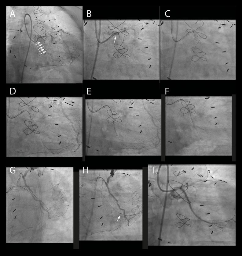
Figure 3.
Case #2. (A) Contrast injection of the left main and the patent LAD graft showed a long, heavily calcified CTO lesion of the proximal CX, with a tapered proximal cap and a steep 90° bend proximal to the CTO lesion (arrows). (B) Puncture of the proximal CTO cap was performed using a balloon-trapped Corsair (Asahi Intecc Co.) microcatheter (arrow). (C) A controlled dissection was created by knuckling of the Fielder XT-A guidewire (Asahi Intecc Co.). (D) The CrossBoss catheter could not advance at the height of the CTO body due to heavy calcification. (E) The Fielder XT-A guidewire was knuckled further down to overcome the calcification. (F) To limit the size of the distal target zone, the CrossBoss catheter was used to cross the last part. (G) Multiple balloon inflations were required to overcome the friction, needed to successfully deliver the Stingray LP catheter. (H) Successful ‘stick-and-swap’ re-entry was performed using the Stingray and Pilot 200 (Abbott Vascular) guidewires (arrow). (I) The native CX was stented without any complications.

