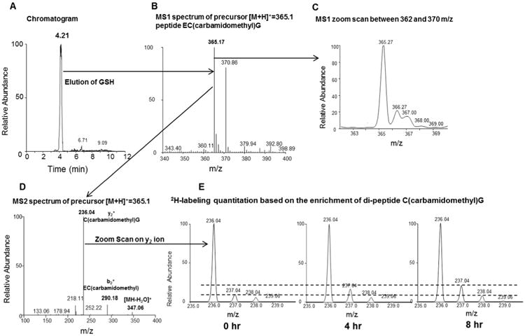
Fig. 2.

Glutathione analysis by LC–MS/MS. Panel A: Carbamidomethylated glutathione derivative is eluted at 4.2 min. Panel B: MS1 spectrum of precursor ion of carbamidomethylated glutathione with m/z 365.15 in positive ion mode. Panel C: MS1 zoom scan of precursor ion monitored between 362 m/z and 370 m/z. Panel D: Collision-indiced dissociation of derivatized glutathione yields abundant cysteine(carbamidomethyl)glycine ion at m/z 236.04 along with other fragment ions. Panel E: The zoom scanned spectra of cysteine(cabamidomethyl)glycine fragment ion before and after (4 h and 8 h) of 2H2O treatment. The dashed lines are added to aid in visualizing differences in the isotopic abundance.
