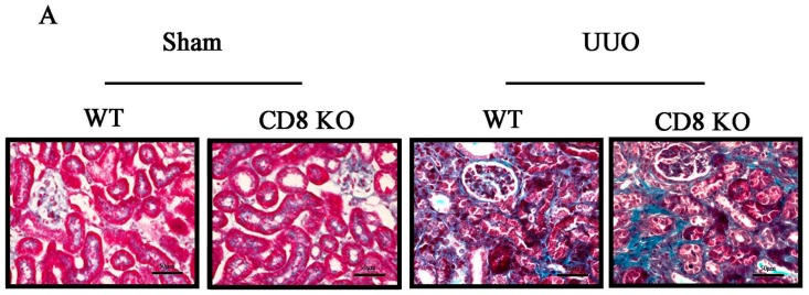
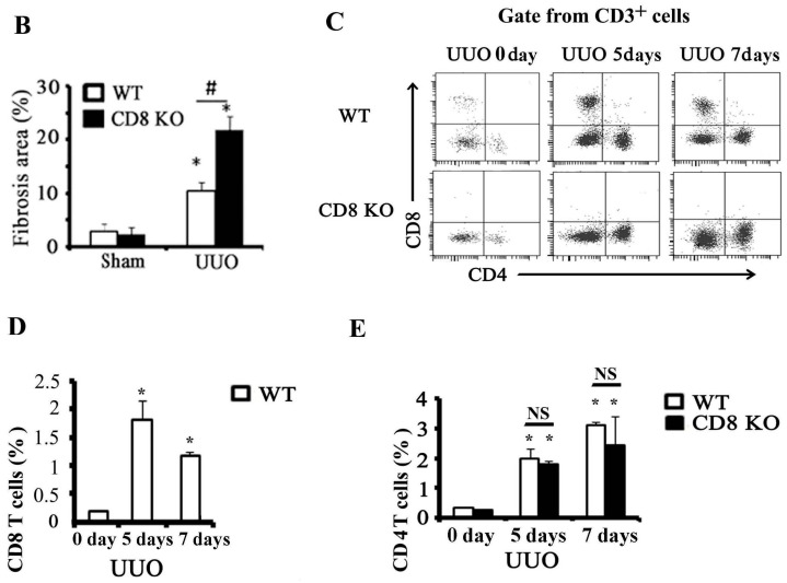
Figure 1.
CD8 deficiency promotes renal fibrosis in unilateral ureteric obstruction (UUO) mice. (A) Masson’s Trichrome staining was performed to examine fibrosis at day 7; (B) Quantitative analysis of fibrosis area in UUO kidneys. CD8 knockout (KO) increases fibrosis in UUO kidney (* p < 0.05 vs. sham, # p < 0.05 vs. wild-type (WT) UUO; n = 5/group). Scale bars, 50 µm; (C) In fluorescence-activated cell sorting analysis, kidney CD8+ T cells were stained with anti-CD45-PerCP-Cy5.5, anti-CD3e-PE-Cy594, anti-CD8-APC-Cy7, and anti-CD4-PE to confirm the absence of CD8+ cells in CD8 KO mice and quantitate (D) CD8+ T cells or (E) CD4+ T cells. (* p < 0.05 vs. WT UUO 0 day, NS: no significant difference; n = 6/group).


