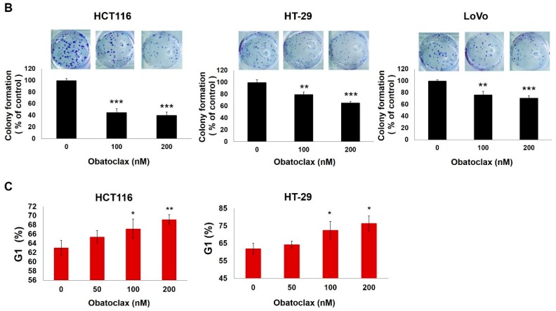
Figure 1.
Antiproliferative action of obatoclax on a panel of human colorectal cancer cell lines. (A) Obatoclax inhibits colorectal cancer cell proliferation. Human colorectal carcinoma cell lines HCT116, HT-29, and LoVo cells (4 × 10⁴) were treated with obatoclax (0, 50, 100, 200 nM) for 24, 48, and 72 h. The numbers of cells at each time point were counted using a Neubauer chamber. Data were presented as the percentage relative to the drug-untreated control; (B) Obatoclax suppresses colony formation by colorectal cancer cells. (Upper panel) HCT116, HT-29, and LoVo cells (2 × 10²) treated with obatoclax (0, 100, 200 nM) for 24 h were allowed to proliferate in drug-free culture media for 10~14 days to form colonies, followed by crystal violet staining for scoring colonies. Shown here are the representative images from three independent experiments. (Lower panel) Quantitative results of the upper panels. Data were presented as the percentage relative to the drug-untreated control; (C) Obatoclax arrests cell cycle progression at the G1-phase. HCT116 and HT-29 cells were treated with obatoclax (0, 50, 100, 200 nM) for 24 h, followed by flow cytometry analyses to determine the levels of cell population at the G1-phase of the cell cycle. Data were presented as the percentage relative to the drug-untreated control. *: p < 0.05; **: p < 0.01; ***: p < 0.001.


