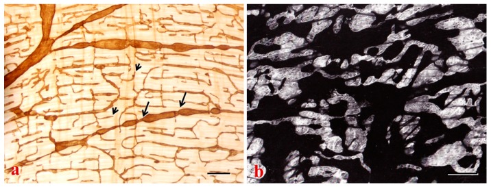
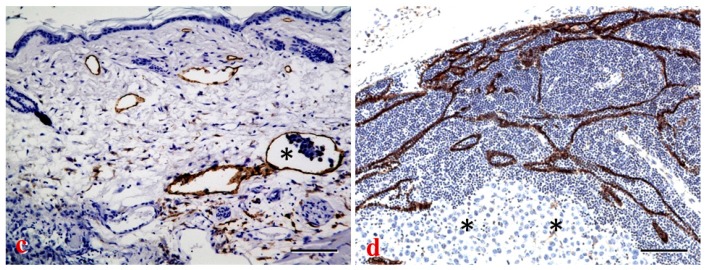
Figure 3.
Lymphatics in normal and tumor tissues. (a,b) Lymphatic networks with 5′-Nase staining are featured by blind ends (arrowheads) and valves (arrows) in the intermuscular layer of the jejunum (a) and the pleural membrane (b), backscattered electron imaging in scanning electron microscopy) of monkeys; (c,d) In the mouse melanoma model, lymphatic vessel endothelial hyaluronan receptor (LYVE-1) staining shows increased lymphatic vessels in the skin (c), and increased subcapsular and cortical lymphatic sinuses in the lymph node (d). The metastatic cells invade lymphatic vessels in the subdermal tissue ((c), asterisk) and aggregate in the LN parenchyma ((d), asterisks). Bars: (a) 500 μm; (b) 150 μm; (c,d) 100 μm.


