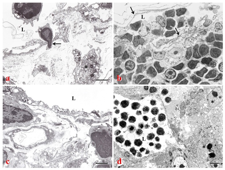
Figure 4.
Peripheral lymphatic vessels (a) and intranodal lymphatic sinuses (b–d) in mouse tissues. (a) In the non-obese diabetic (NOD) pancreas, a lymphocyte is penetrating the lymphatic vessel stained with 5′-Nase cerium (arrow). The asterisk indicates a macrophage; (b) In the LN of Bagg albino/c (BALB/c) mice, the subcapsular sinus is filled with lymphocytes and DCs. The arrows indicate endothelial layers of the lymphatic sinus; (c) The medullary sinus of NOD mice is decorated with 5′-Nase cerium particles, and metabolic products of inflamed cells are seen beneath the endothelial layer; (d) The cortical sinus filled with lymphocytes is surrounded by metastatic melanoma cells (asterisks). L: lymphatic vessels or sinuses. Bars: (a,c) 2 μm; (b,d) 5 μm.

