Figure 2.
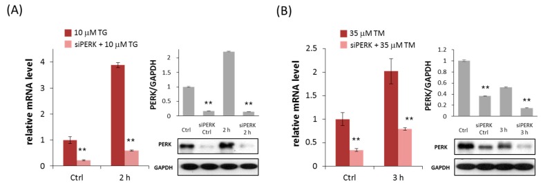
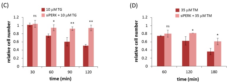
PERK silencing increases cell viability during persistent ER stress. The efficiency of PERK silencing was checked both on mRNA (left panel) and protein (right panel) levels followed in time via (A) TG (10 µM) and (B) TM (35 µM) treatment. The mRNA level was followed by real-time PCR and the expression level of PERK was followed by Western blot with/without addition of PERK siRNA for 2 (TG) and 3 h (TM) long treatment. GAPDH was used as housekeeping gene. The intensity of PERK is normalized for GAPDH. The amount of viable cells was assessed in PERK-silenced cells after (C) TG (10 µM) or (D) TM (35 µM) treatment in time. The amount of viable HEK293T cells was followed in time by measuring the percentage of cells permeable to trypan blue. Three parallel experiments were carried out and the amount of viable cells (lower panel) was plotted (errors bars represent standard deviation, asterisks indicate statistically significant difference: * p < 0.05; ** p < 0.01); ns: not significant.


